Expert Eye Care in Cyprus
Dr Stavroula Chrysostomou Kaponidi
Consultant Ophthalmic Surgeon — Nicosia & Paphos, Cyprus

MD, CCST in Ophthalmology
University of Patras, Greece
9+ Years Experience
Specialist ophthalmic care
International Training
Athens & Nicosia hospitals
Patient-Centered
Evidence-based approach
 Registered GESY Healthcare Provider
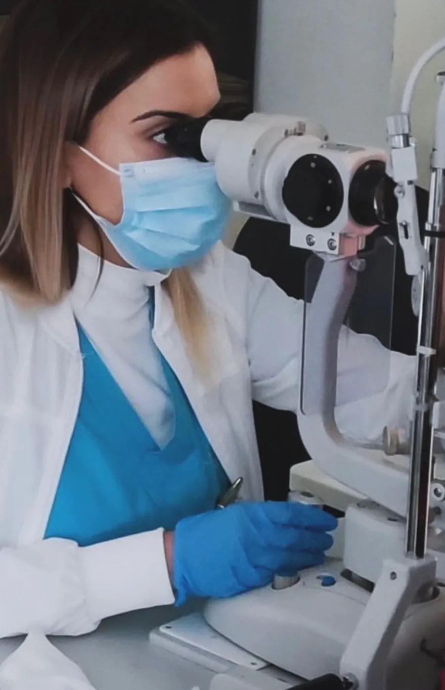
Dedicated to Your Vision
Dr Stavroula Chrysostomou Kaponidi is a highly qualified Consultant Ophthalmic Surgeon with extensive experience in comprehensive eye care, surgical treatment, and medical retina.
She holds an MD from the University of Patras, Greece, and a Certificate of Completion of Specialist Training (CCST) in Ophthalmology. With over 9 years of specialized experience, Dr Chrysostomou Kaponidi has worked at leading institutions including Attikon University Hospital Athens, Archbishop Makarios III Hospital Nicosia, and Nicosia General Hospital.
Her practice is built on a patient-centered approach and evidence-based care, ensuring each patient receives personalized treatment tailored to their specific needs. Dr Chrysostomou Kaponidi regularly participates in national and international ophthalmology congresses to stay at the forefront of medical advances.
Areas of Expertise
Comprehensive ophthalmic care using the latest evidence-based treatments and state-of-the-art technology.
Cataract Surgery
Modern cataract surgery with intraocular lens implantation
Glaucoma Diagnosis & Management
Detection and treatment of all types of glaucoma
Medical Retina & Macular Diseases
Specialist care for retinal and macular conditions
Intravitreal Injections
Treatment of AMD, diabetic macular oedema and retinal vascular diseases
Refractive Laser Surgery
Advanced laser correction of myopia, hyperopia and astigmatism
Keratoconus & Corneal Cross-Linking (CXL)
Assessment and management including CXL when indicated
Diabetic Eye Disease
Screening, monitoring and treatment of diabetic retinopathy
Comprehensive Ophthalmic Examinations
Full eye assessments using state-of-the-art diagnostic technology
24/7 Emergency
Emergency Ophthalmology
Urgent management of acute eye conditions with accuracy and confidence
First Appointment
Patient Medical & Ophthalmic History Form
What information will I be asked for?
We will ask about your general health conditions, current medications, allergies, previous surgeries, and family history of eye disease. For your ophthalmic history, we will ask about any previous eye conditions, eye surgeries, current eye medications, when you last had your eyes checked, and any changes you have noticed in your vision.
Why is a complete history important?
A thorough medical and ophthalmic history helps us provide the safest and most effective care. Many general health conditions and medications can affect your eyes and influence treatment decisions. For example, diabetes affects the retina, autoimmune conditions can cause inflammation in the eye, and blood thinning medications are relevant if surgery is being considered.
What should I bring to my first appointment?
Please bring a list of your current medications, any relevant medical or ophthalmic reports, your current glasses or contact lenses, and a list of any allergies. If you have had previous eye tests or imaging, please bring the results or ask your previous provider to share them.
Patient Information
Comprehensive information about common eye conditions, treatments, and what to expect during your visit.
Open Angle Glaucoma
Open angle glaucoma is the most common type of glaucoma. It happens when the eye's drainage system gradually becomes less efficient, causing fluid to build up and increase the pressure inside your eye. This elevated pressure can slowly damage the optic nerve, which carries visual information from your eye to your brain.
Open angle glaucoma is often called the "silent thief of sight" because in its early stages, there are usually no symptoms at all. You might not notice any vision loss until the disease has already caused significant damage. By the time you notice problems, typically with peripheral (side) vision, substantial irreversible damage may have occurred. This is why regular eye examinations are crucial.
Based on the latest research, we recommend starting with a laser treatment called Selective Laser Trabeculoplasty (SLT). This is now considered the gold standard first-line treatment for open angle glaucoma. It's a quick, safe office procedure that lowers your eye pressure without the need for daily eye drops. If we need additional pressure lowering later, we can add medications or consider other procedures.
SLT stands for Selective Laser Trabeculoplasty. It targets the drainage system in your eye to help fluid flow out more easily, which lowers your eye pressure. It's called "selective" because it only affects specific cells without damaging the surrounding tissue, which means it's very safe and can even be repeated if needed years later. The entire procedure takes about 5 to 10 minutes.
Recent large studies, particularly the LiGHT trial, have shown that patients who start with SLT actually do better than those who start with eye drops. About 70% of patients who have SLT remain medication-free for years, avoiding the side effects and daily hassle of drops. SLT patients also showed 29% slower disease progression and needed fewer surgeries compared to patients who started with drops.
For most patients, the pressure-lowering effect of SLT lasts 2 to 3 years, and sometimes even longer. The great thing is that if your pressure starts to rise again, we can safely repeat the procedure. About 70% of patients remain at their target pressure without needing drops or surgery for at least 3 years.
The procedure is done in the clinic. We place numbing eye drops so you won't feel pain. You'll sit at a machine similar to the one used for regular eye exams, and a special lens is placed on your eye. The laser treatment takes about 5 to 10 minutes. You may see some flashes of green light during the procedure. Afterward, we'll use anti-inflammatory drops and check your pressure. Most patients can return to normal activities the same day.
After SLT, we'll want to see you within the first few weeks to check how your pressure is responding. Once we confirm the treatment is working, we typically monitor you every few months initially, then can often space visits to every 6 months or annually, depending on how stable your pressure remains.
Angle Closure Glaucoma
Angle closure glaucoma occurs when the drainage angle of the eye becomes partially or completely blocked. This can lead to a sudden or gradual rise in eye pressure, which may damage the optic nerve.
Acute angle closure is a sudden blockage causing rapid pressure rise, severe pain, and urgent symptoms. Chronic angle closure develops gradually, with the angle slowly closing over time. Both forms require treatment.
Symptoms include sudden, severe eye pain, headache, nausea or vomiting, blurred vision, seeing halos around lights, and a red eye. These symptoms require immediate medical attention, as untreated acute angle closure can cause permanent vision loss within hours.
YAG Laser PI is a preventive laser procedure where a tiny hole is made in the iris to improve fluid flow within the eye and prevent angle closure. It is the standard first-line treatment for angle closure and is usually performed as a preventive measure in at-risk eyes.
The procedure is quick and done in the clinic. Numbing drops are applied, and a special contact lens is placed on the eye. The YAG laser is used to create a small opening in the iris. Most patients feel only a mild sensation. The entire procedure takes just a few minutes.
YAG PI is generally very safe. Possible but uncommon side effects include temporary blurred vision, mild inflammation, a small rise in eye pressure, or minor bleeding at the laser site. Serious complications are rare.
After the procedure, your vision may be slightly blurry for a short time, and you may notice a mild ache. Anti-inflammatory drops are typically prescribed for a few days. You will usually have a follow-up visit within a few weeks to check the iridotomy and your eye pressure.
Cataract
A cataract is a condition where the natural lens of the eye becomes cloudy. This causes blurred or reduced vision.
• Aging (most common cause) • Diabetes • Long-term sunlight exposure • Eye injury • Smoking • Certain medications
• Blurred or cloudy vision • Difficulty seeing at night • Sensitivity to bright light • Faded or dull colours • Seeing halos around lights • Frequent change of glasses
No. Cataract usually causes no pain.
Vision will slowly get worse. Daily activities such as reading, driving, or recognising faces may become difficult. In advanced cases, severe vision loss or blindness can occur. Advanced cataract can lead to increased eye pressure, eye inflammation, and may make surgery more difficult later.
Cataract is treated with surgery. There are no eye drops or medicines that can remove a cataract.
Cataract surgery is a safe and common procedure. The cloudy lens is removed and replaced with a clear artificial lens. The procedure is performed using ultrasound to break up and remove the cloudy lens.
Yes. Cataract surgery has a very high success rate.
In most cases, one surgery is enough. In rare situations, a second surgery may be needed due to complications or lens movement.
Most patients recover well. Rare complications may include: • Infection • Inflammation or redness • Swelling • Increased eye pressure • Blurred vision • Sensitivity to light
• Use eye drops exactly as prescribed • Do not rub the eye • Keep the eye clean • Wear eye protection • Avoid heavy lifting and bending • Attend follow-up appointments
• Severe eye pain • Sudden loss of vision • Increasing redness or discharge • Flashes of light
Most people see improvement within a few days. Complete healing may take a few weeks.
Keratoconus
Keratoconus is a progressive eye condition in which the cornea (the clear front surface of the eye) gradually thins and bulges outward into a cone shape. This irregular shape distorts vision and can make everyday tasks like reading and driving difficult.
The exact cause is not fully understood, but it is believed to involve a combination of genetic, environmental, and hormonal factors. Frequent and vigorous eye rubbing is strongly associated with keratoconus progression and should be avoided. It often begins during the teenage years or early twenties and may progress for 10 to 20 years before stabilizing.
In the early stages, keratoconus may cause slight blurring and distortion of vision, increased sensitivity to light and glare, and frequent changes in spectacle prescription. As the condition progresses, vision may become significantly distorted, and standard glasses may no longer correct it adequately.
Treatment depends on the stage. In early stages, glasses or soft contact lenses may correct the vision. As it progresses, rigid gas-permeable or scleral contact lenses may be needed. To slow or halt progression, corneal collagen cross-linking (CXL) is a very effective treatment.
CXL is a minimally invasive procedure designed to strengthen the cornea and prevent further thinning and bulging. It involves applying riboflavin (vitamin B2) drops to the cornea and then exposing it to a controlled dose of ultraviolet A (UVA) light. This process strengthens the chemical bonds (cross-links) between the collagen fibres in the cornea.
The procedure itself is not painful, as the eye is numbed with anaesthetic drops. After the procedure, you may experience some discomfort, tearing, and sensitivity to light for a few days. Pain relief is usually managed with oral painkillers and lubricating eye drops.
The primary goal of CXL is to halt (stop) the progression of keratoconus, not necessarily to improve your current vision. In many cases, vision does stabilize and some patients even experience a modest improvement in the months following the procedure. Contact lenses or glasses may still be needed for optimal vision correction.
After the procedure, a bandage contact lens is placed on the eye and is usually removed after 5 to 7 days. You will need to use antibiotic and anti-inflammatory eye drops as prescribed. Most patients return to work within a week. Full visual recovery and corneal stabilization may take several weeks to months.
Macular Degeneration (Dry)
Dry macular degeneration (also called dry AMD) is a common age-related condition that affects the macula, the central part of the retina responsible for sharp, detailed vision. It is characterized by the gradual breakdown and thinning of the macular tissue, often with the appearance of small deposits called drusen.
Symptoms may develop gradually and include blurred or reduced central vision, difficulty reading or recognising faces, needing brighter light for close tasks, increased difficulty adapting to low light, and a gradual increase in the haziness of overall vision. A key symptom is that straight lines may start to appear wavy or distorted.
The exact cause is not fully understood, but it is strongly associated with aging. Risk factors include age (over 50), family history, smoking, high blood pressure, obesity, and prolonged sun exposure. Genetics also play a significant role.
There is currently no cure for dry AMD, but its progression can potentially be slowed. The AREDS2 (Age-Related Eye Disease Study 2) formula of nutritional supplements has been shown to reduce the risk of progression in intermediate-stage dry AMD. A healthy diet rich in leafy greens and fish, regular exercise, not smoking, and protecting your eyes from UV light are also recommended.
If you notice any sudden changes in your vision, particularly if straight lines appear wavy or distorted, or if there is a rapid decline in central vision, you should seek urgent assessment. These symptoms could indicate a conversion from dry to wet AMD, which requires prompt treatment. Regular monitoring with an Amsler grid at home is recommended.
Wet Macular Degeneration
Wet macular degeneration (also called wet AMD or neovascular AMD) is a more advanced and serious form of age-related macular degeneration. It occurs when abnormal blood vessels grow underneath the retina and leak fluid or blood, causing rapid damage to the macula.
The main treatment for wet AMD is anti-VEGF (vascular endothelial growth factor) therapy, which involves regular intravitreal injections into the eye. These medications help to stop the growth of abnormal blood vessels and reduce leakage, stabilizing or improving vision in many patients. Prompt treatment is essential to prevent irreversible vision loss.
Intravitreal injections are a well-established, safe procedure where medication is injected directly into the vitreous (the gel-like substance inside the eye). The eye is numbed with anaesthetic drops, and the injection is performed under sterile conditions. The procedure takes only a few minutes and most patients experience only a mild sensation of pressure.
Treatment usually begins with a course of monthly injections for the first 3 months (a "loading phase"). After that, the frequency of injections is individualized based on your response. Some patients may require injections every 4 to 8 weeks, while others may be able to extend intervals. Regular monitoring with OCT scans helps guide the treatment schedule.
Diabetic Retinopathy
Diabetic retinopathy is a complication of diabetes that affects the blood vessels of the retina. Over time, high blood sugar levels can damage the small blood vessels in the retina, causing them to leak fluid, bleed, or become blocked. This can lead to vision impairment or blindness if left untreated.
There are four main stages: mild non-proliferative (small areas of swelling in retinal blood vessels), moderate non-proliferative (some blood vessels become blocked), severe non-proliferative (many more vessels are blocked, depriving areas of the retina of blood supply), and proliferative diabetic retinopathy (the most advanced stage where new, abnormal blood vessels grow on the retina).
Treatment depends on the stage and severity. In the early stages, careful monitoring and good control of blood sugar, blood pressure, and cholesterol may be all that is needed. For diabetic macular oedema, anti-VEGF intravitreal injections and in some cases laser treatment are used. For proliferative diabetic retinopathy, laser treatment (panretinal photocoagulation) or vitrectomy surgery may be required.
Good control of your blood sugar levels is the most important factor. Also important are controlling blood pressure and cholesterol, having regular dilated eye examinations, not smoking, and maintaining a healthy lifestyle. Annual eye screening is recommended for all people with diabetes.
Astigmatism
Astigmatism is a common refractive error caused by an irregularly shaped cornea or lens. Instead of being evenly round (like a football), the cornea is shaped more like a rugby ball, causing light to focus unevenly on the retina. This results in blurred or distorted vision at all distances.
Astigmatism can be corrected with glasses (using cylindrical lenses), toric contact lenses, or refractive laser surgery such as LASIK or PRK. During cataract surgery, a toric intraocular lens can be used to correct astigmatism simultaneously. The most suitable option depends on the degree of astigmatism yet also other eye conditions.
Symptoms include blurry or distorted vision at all distances, difficulty seeing at night, eye strain, squinting, headaches (especially after prolonged reading or screen work), and difficulty seeing fine details. Many people have mild astigmatism without noticeable symptoms.
Astigmatism can change over time, sometimes increasing gradually. In most cases, it is a stable condition. However, a significant or rapid increase in astigmatism, especially if associated with distorted vision, should be evaluated, as it may indicate keratoconus or other corneal conditions.
Posterior Vitreous Detachment (PVD)
Posterior vitreous detachment (PVD) is a common eye condition that usually happens as part of normal aging. Inside the eye there is a clear, jelly-like substance called the vitreous gel. The vitreous fills the middle of the eye and is attached to the retina, the light-sensitive tissue at the back of the eye. As people get older, the vitreous becomes more liquid and can pull away from the retina. This separation is called a posterior vitreous detachment. PVD is very common after the age of 50 and often does not threaten vision. However, in some cases, the pulling of the vitreous can create a retinal tear or retinal detachment, which requires urgent treatment.
You may notice: • Sudden appearance of floaters (small black spots, cobwebs, threads, or moving shadows) • Flashes of light, especially in dim lighting or side vision • Temporarily blurred or hazy vision • A sensation of movement inside the eye Symptoms are often more noticeable during the first few days or weeks.
Most cases of PVD are harmless and settle without treatment. However, a small number of patients may develop: • A retinal tear • A retinal detachment • Bleeding inside the eye These complications can permanently affect vision if not treated quickly.
Contact an eye doctor or emergency eye clinic immediately if you develop: • A sudden increase in floaters • A shower of black spots • Persistent flashes of light • A dark curtain or shadow in your vision • Loss of side vision • A sudden decrease in vision These symptoms may indicate a retinal tear or retinal detachment.
Risk factors include: • Aging • Nearsightedness (myopia) • Previous eye surgery (such as cataract surgery) • Eye injury • Diabetes • Inflammation inside the eye
An eye specialist examines the retina using: • A dilated eye examination • A slit lamp examination • Sometimes ultrasound imaging The purpose of the examination is mainly to rule out retinal tears or retinal detachment.
In most patients: • No treatment is needed • Floaters usually become less noticeable over weeks to months • Flashes generally decrease with time Surgery is rarely required unless serious complications develop.
The first several weeks are the most important, because retinal tears are more likely to occur during this period. Recommended: • Attend all follow-up appointments • Monitor your symptoms carefully • Rest your eyes if they feel strained • Stay hydrated and maintain good general health Usually safe: • Reading • Watching television • Using a computer or phone • Gentle walking and normal daily activities Discuss with your doctor before: • Heavy lifting • High-impact exercise • Contact sports • Activities involving sudden head movement
Floaters can be annoying at first, but the brain often adapts over time. Many patients notice significant improvement within a few months. Helpful tips: • Move your eyes gently up and down to shift floaters away from central vision • Use good lighting for reading • Wear sunglasses outdoors if flashes are bothersome
Yes. Even if symptoms improve, follow-up examinations are important because retinal tears can occasionally appear later. Typical follow-up may occur: • Within several weeks after diagnosis • Earlier if symptoms worsen
Most people with PVD maintain normal vision and recover without complications. Prompt medical attention for warning symptoms greatly reduces the risk of permanent vision loss. Seek urgent ophthalmology review immediately if you notice: • New flashes • More floaters • A curtain or shadow in your vision • Sudden visual loss Early treatment of retinal tears can protect your eyesight.
Clinic Locations
Dr Chrysostomou Kaponidi provides comprehensive eye care at three locations across Cyprus.
The map is provided by Google. Loading it shares data with Google. Privacy Policy
Nicosia Clinic
6 Grigori Afxentiou St, Kokkinotrimithia 2660
Nicosia, Cyprus
Get DirectionsThe map is provided by Google. Loading it shares data with Google. Privacy Policy
Paphos — Saint George Private Hospital
29 Eleftheriou Venizelou Street
Paphos 8021, Cyprus
Get DirectionsThe map is provided by Google. Loading it shares data with Google. Privacy Policy
Paphos — WeCare Medical Centre
14 Mesogis Avenue, Mesogi
Paphos 8280, Cyprus
Get DirectionsContact Us
Contact us today for any questions about our services or to learn more about how we can help you.